Skip to content

Frequency Foundation

Twice the Energy with Half the Stress

Carcinogenesis: Preventing it with electronic medicine

Update: This original Rife organism appears to be a necessary condition for cancer cells to survive. Eliminating this organism causes cancer cell death. There are many strains of this organism and it is very persistent.
[trx_button type=”square” style=”filled” size=”small” align=”left” link=”https://www.frequencyfoundation.com/product/original-rife-frequencies/” popup=”no” top=”inherit” bottom=”inherit” left=”inherit” right=”inherit”]Original Rife Frequencies[/trx_button]

As medical research advances, the strategy of systematically eliminating cancer cells with electromagnetic frequencies is entering the mainstream. A recent clinical trial showed clearly that low voltage application of frequencies destroys cancer cells, although their approach would need significant modification to be fully effective. Clinical trials have begun and electromagnetic frequencies have been approved by the FDA to treat certain tumor types. A remarkable video by Bill Doyle shows frequencies disrupting growth of cancer cells.

There are several steps to  dealing with cancer cells:
1. Nutritional and lifestyle factors described elsewhere are critical
2. Eliminate the Rife BX BY complex
3. Eliminate the Gregory cancer virus
4. Eliminate parasites that promote tumor growth
5. Disrupt glucose metabolism of cancer cells
6. Eliminate viruses that cause malignant cells
7. Target specific malignant cells for elimination

——
There is abundant epidemiological data from all over the world on cancer incidence for a wide variety of tumors in animal and human populations. My thesis advisor was Editor of the Journal of the National Cancer Institute in the late 1970s and early 1980s. He gave me a stack of over 300 papers from the medical journals with carcinogenesis dose reponse curves for many different species. All the curves looked different. He told me to explain why they were all different and he would give me a Ph.D. I then spent about 8 years doing supercomputer mathematical analysis of data on various types of cancer. The final data set I focused in on for publication was data from the Third National Cancer Survey which had complete coverage from many states in the U.S. and was the best available data at the time. My thesis advisor had led the survey and I had complete access to all data at the National Cancer Institute. The core model we developed is now the generally accepted model for carcinogensis. It was initially validated for colon cancer. See:
Sutherland, JV and Bailar, JC. The multihit model of carcinogenesis: etiologic implications for colon cancer. J Chronic Dis. 1984;37(6):465-80.

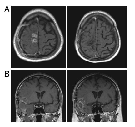
For most human cancers, the data clearly shows a four hit process. This can be visualized most clearly in skin tumors. An abnormal cell appears and begins dividing and creates a small patch on the skin. Within this lesion, a cell mutates, and you have a small patch growing within a patch. This happens a third time, then a fourth time. On the fourth genetic change, the cell breaks through the mechanisms that control proliferation and is malignant. It grows uncontrollably in a fifth phase, with the right promoting environment available, and can get into the bloodstream and migrate to other parts of the body causing metastases.

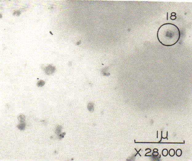
I have repeatedly seen the frequencies 2008 and 2127 appear in myself and others after eating certain food. I have been able to confirm that this has happened in most people who have eaten a specific meal. This is the BX and BY “virus” form of Rife’s organism. Today, this is not believed to be a virus and some think it is a mycoplasma. Recent DNA sequencing of organisms found in cancer cells showed them to be a fungus.

These organisms are clearly not a typical virus. They immediately go systemic throughout the entire body. And it originally took days of repeated treatment for long periods with the FSCAN and/or EM6+ to clear the body of them. With newer technologies they can be eliminate a lot faster.

However, more and more evidence is appearing that shows cancer cell frequencies are specific frequencies in a virus sequence. It appears that a virus disrupting the cellular machinery is one of the critical steps (but not the only step) that produces a tumor. Therefore, eliminating the relevant viruses is critical to prevent ongoing appearance of pre-malignant or malignant cells.

I have repeatedly seen non-malignant skin tumors grow quickly and in one case apparently become malignant in the presence of BX and BY. In the presence of this organism it is common to have tumors erupt in less than a day. This helps to explain an interesting aspect of my previous research in carcinogenesis. In some tumors, prostate for example, the data show that the first malignant cell appears 35 years before clinical diagnosis on the average. In a fast growing tumor like lung cancer, the first malignant cell appears about a year before clinical diagnosis. However, it is very common to see people go downhill extremely quickly after initial diagnosis. While some of this is undoubtedly due to the shock of the diagnosis and resultant depression of the immune system, it is aggrevated by the BX and BY virus. Much of conventional cancer treatment does not eliminate the virus and may even promote its growth.

So the BX and BY organisms are strong promoters of at least stage 3 (premalignant) and stage 4 (malignant) tumors and may even promote earlier stages. As indicated in the paper by Owens DM, Wei S, Smart RC. A multihit, multistage model of chemical carcinogenesis. Carcinogenesis, Vol. 20, No. 9, 1837-1844, September 1999, promotion of early phases of tumor initiation increases the cellular population at risk of mutating into a subsequent phase. This process happens so fast in the presence of BX and BY that I believe the Rife organisms are both promoters and mutagens.

The first approach to stopping proliferation of tumors must be to eradicate the BX and BY virus. Parasites also release substances that promote tumor growth so they must be eliminated as well. However, they are not as dangerous as BX and BY. I believe if everyone was monitored for BX and BY and the infection was eliminated immediately, we could significantly cut cancer incidence in the U.S., probably by more than 50%.

The fact that this information has been available for almost 100 years now shows how closed mindedness and suppression of innovation has compromised American medicine. This phenomenon has largely been driven by business interests, initially by a director of the AMA who was ultimately convicted of fraud and conspiracy for suppressing cancer therapies, and in more recent years by the pharmaceutical industry.

The tumors I have worked with (lung, cervix, skin, colon) have all had the presence of BX and BY which can be eliminated straight away. It is important to realize when targeting the BX and BY organisms with an electronic device that when you kill one strain, another strain proliferates and the frequency changes. These organisms have very stable frequencies around 2008 and 2127, however, and I have seen them deviate only by 3HZ at the most. It is important to eliminate all of them and you must have the exact frequency to 1HZ and this frequency may vary between 2003-2010 and 2125-2130. In recent years, the frequency set for BX BY has expanded to hundreds of frequencies.

Eliminating the BX and BY organism does not stop cells which have already reached stage 4 and are malignant. In fact, all malignant tumors will not show BX and BY present. They can continue to proliferate and metastasize without BX and BY. The best way to eliminate the malignant cells is to stop the ATP metabolism in these cells while leaving normal cells unaffected. This will prevent mitosis and the cancer cells will die a normal death without proliferating. This appears to be possible with frequencies in the 11,700,000 range. These frequencies also seem to stop mitosis of stage 3 cells as well. The data in the Gorgun paper shows how this is possible.

Gorgun SS. Studies on the Interaction Between Electromagnetic
Fields and Living Matter Neoplastic Cellular Culture. Frontier Perspectives 7:2:44-59, Fall 1998.

Stopping cellular mitosis requires the exact frequency. I have seen frequencies in the range 11,600,000 – 11,900,000HZ affect tumors. Different tumors of the same type (or perhaps cells in different stages) will have slightly different frequencies. Metastatic tumors will have different frequencies. They must all be eliminated systematically, one by one.

I believe that Rife was able to affect a “cure” in almost every case because his device running at 11,700,000 had enough variability and harmonics to stop mitosis in all relevant cells, even though their frequencies may have been slightly different. The curse of modern technology is that it is so precise, most people are unable to reproduce Rife’s work.

Richard Loyd had a machine some years ago that could treat directly at 11,700,000HZ. Most of us are not so lucky. I have found some success, however, in using the FSCAN to treat all the octaves of the exact frequencies in the FSCAN range from 10-3,000,000HZ. More recent FSCAN devices work in the 11-12MHZ range. Current frequency sets provided to Frequency Foundation subscribers go well over 30MHZ to target viral patterns in the bodies energy field. It will be difficult but not impossible, in my view, to stop all cellular mitosis using a Rife device that can only treat at less than 10,000HZ.

These comments represent a working hypothesis that has been successful in many cases and not as successful in rapidly growing tumors. It needs further research and new data may alter the working hypothesis. I present it so that others can take a look and see if they can produce the same results consistently.

Milkweed Cures Skin Cancer

The sap from Euphorbia peplus is effective against human nonmelanoma skin cancers

  1. J.R. Ramsay1
  2. A. Suhrbier2,3
  3. J.H. Aylward4,
  4. S. Ogbourne2
  5. S.-J. Cozzi2
  6. M.G. Poulsen1,
  7. K.C. Baumann1
  8. P. Welburn4
  9. G.L. Redlich4,
  10. P.G. Parsons2,3
Article first published online: 27 JAN 2011
DOI: 10.1111/j.1365-2133.2010.10184.x
© 2011 The Authors. BJD © 2011 British Association of Dermatologists 2011
Issue
British Journal of Dermatology

British Journal of Dermatology

Early View (Articles online in advance of print)
Additional Information(Show All)
  1. Funding sources This work was conducted with financial assistance from Peplin Biotech Pty Ltd and an Australian Commonwealth Government Industry Research and Development Board R&D START grant.
  2. Conflicts of interest J.H.A. and G.L.R. were, and P.W. and S.O. are currently, employees of Peplin Inc. (formerly Peplin Biotech Pty Ltd). J.R.R., A.S. and P.G.P. were consultants to Peplin Biotech Pty Ltd. Peplin Inc. is now wholly owned by LEO Pharma.

Summary

Background  The sap from Euphorbia peplus, commonly known as petty spurge in the U.K. or radium weed in Australia, has been used as a traditional treatment for a number of cancers.
Objective  To determine the effectiveness of Epeplus sap in a phase I/II clinical study for the topical treatment of basal cell carcinomas (BCC), squamous cell carcinomas (SCC) and intraepidermal carcinomas (IEC).
Methods  Thirty-six patients, who had refused, failed or were unsuitable for conventional treatment, were enrolled in a phase I/II clinical study. A total of 48 skin cancer lesions were treated topically with 100–300 μL of E. peplus sap once daily for 3 days.
Results  The complete clinical response rates at 1 month were 82% (= 28) for BCC, 94% (= 16) for IEC and 75% (= 4) for SCC. After a mean follow-up of 15 months these rates were 57%, 75% and 50%, respectively. For superficial lesions < 16 mm, the response rates after follow-up were 100% for IEC (= 10) and 78% for BCC (= 9).
Conclusions  The clinical responses for these relatively unfavourable lesions (43% had failed previous treatments, 35% were situated in the head and neck region and 30% were > 2 cm in diameter), are comparable with existing nonsurgical treatments. An active ingredient ofE. peplus sap has been identified as ingenol mebutate (PEP005). This clinical study affirms community experience with E. peplus sap, and supports further clinical development of PEP005 for the treatment of BCC, SCC and IEC.

Cancer clinical trial shows good response from frequencies

J Exp Clin Cancer Res. 2009 Apr 14;28:51.

Amplitude-modulated electromagnetic fields for the treatment of cancer: discovery of tumor-specific frequencies and assessment of a novel therapeutic approach.
Cabinet Médical, Avenue de la Gare 6, Lausanne, Switzerland. [email protected]
Abstract
PURPOSE: Because in vitro studies suggest that low levels of electromagnetic fields may modify cancer cell growth, we hypothesized that systemic delivery of a combination of tumor-specific frequencies may have a therapeutic effect. We undertook this study to identify tumor-specific frequencies and test the feasibility of administering such frequencies to patients with advanced cancer.
PATIENTS AND METHODS: We examined patients with various types of cancer using a noninvasive biofeedback method to identify tumor-specific frequencies. We offered compassionate treatment to some patients with advanced cancer and limited therapeutic options.
RESULTS: We examined a total of 163 patients with a diagnosis of cancer and identified a total of 1524 frequencies ranging from 0.1 Hz to 114 kHz. Most frequencies (57-92%) were specific for a single tumor type. Compassionate treatment with tumor-specific frequencies was offered to 28 patients. Three patients experienced grade 1 fatigue during or immediately after treatment. There were no NCI grade 2, 3 or 4 toxicities. Thirteen patients were evaluable for response. One patient with hormone-refractory breast cancer metastatic to the adrenal gland and bones had a complete response lasting 11 months. One patient with hormone-refractory breast cancer metastatic to liver and bones had a partial response lasting 13.5 months. Four patients had stable disease lasting for +34.1 months (thyroid cancer metastatic to lung), 5.1 months (non-small cell lung cancer), 4.1 months (pancreatic cancer metastatic to liver) and 4.0 months (leiomyosarcoma metastatic to liver).
CONCLUSION: Cancer-related frequencies appear to be tumor-specific and treatment with tumor-specific frequencies is feasible, well tolerated and may have biological efficacy in patients with advanced cancer.
TRIAL REGISTRATION: clinicaltrials.gov identifier NCT00805337.

Cancer Cells have Electrical Switch

An electrical switch for cancer?

A new type of electrically active cell can turn stem cells cancerous from a distance


[Published 19th October 2010 02:21 PM GMT]


Biologists have identified a new cell type in vertebrates that remotely mediates the transformation of stem cells into either healthy skin cells or cancerous melanoma, providing a potential new tool for researchers in both oncology and regeneration biology. 

A model of melanocyte control 
by transmembrane potential of 
remote “instructor cells”.
Image: Disease Models and Mechanisms. 
This figure was taken from an 
Open Access article distributed 
under the terms of the Creative Commons 
Attribution Non-Commercial Share 
Alike License 

In a paper published Tuesday (19 October) in Disease Models and Mechanisms, the researchers, led by Michael Levin of Tufts University, describe how, by manipulating the voltage across the cell membranes of these so-called “instructor cells” in frog tadpoles in vivo, they have been able to dictate the fate of the descendants of neural crest stem cells “with exquisite specificity.” De-polarizing the cells led to aggressive, metastatic melanoma. A similar effect was found in human pigment cells in vitro. 

The instructor cells themselves lie outside of the neural crest, but appear to communicate with the stem cell population via a long-range signal based on serotonin. Levin told The Scientist that, while the idea that the electrical state of cells is involved in the formation of tumors is not a new one, his study shows that the electrical properties of one type of cell can induce other, distant cells to change their behavior, and might be “a key switch that mediates the stem cell-cancer cell distinction.”


Read more: An electrical switch for cancer? – The Scientist – Magazine of the Life Sciences http://www.the-scientist.com/news/display/57754/#ixzz168Rj0CcT

Cancer Treatment by Electromagnetic Fields Goes Mainstream

Before and after applying 200khz frequencies to brain cancer. Disease has stabilized.
Alternating electric fields arrest cell proliferation in animal tumor models and human brain tumors. Proceedings National Academy of Sciences, June 12, 2007 vol. 104 no. 24 10152-10157.
Author Affiliations
  • *NovoCure Limited, Matam Advanced Technology Centre, Haifa 31905, Israel;
  • Na Homolce Hospital, Roentgenova 2, 150 30 Prague 5, Czech Republic;
  • Rambam Medical Center, PO Box 9602, Haifa 31096, Israel;
  • §Basel University Hospitals, Hebelstrasse 32, 4031 Basel, Switzerland;
  • Centre National de la Recherche Scientifique, Laboratoire d’immunologie et Embryologie Moléculaire, Rue de la Ferollerie, 45071 Orleans, France;
  • Weizmann Institute of Science, PO Box 26, Rehovot 76100, Israel; and
  • **B. Rappaport Faculty of Medicine, Technion–Israel Institute of Technology, Technion City, Haifa 32000, Israel
  • Communicated by Joseph Schlessinger, Yale University School of Medicine, New Haven, CT, April 5, 2007 (received for review January 15, 2007) 
Abstract
We have recently shown that low intensity, intermediate frequency, electric fields inhibit by an anti-microtubule mechanism of action, cancerous cell growth in vitro. Using implanted electrodes, these fields were also shown to inhibit the growth of dermal tumors in mice. The present study extends these findings to additional cell lines [human breast carcinoma; MDA-MB-231, and human non-small-cell lung carcinoma (H1299)] and to animal tumor models (intradermal B16F1 melanoma and intracranial F-98 glioma) using external insulated electrodes. These findings led to the initiation of a pilot clinical trial of the effects of TTFields in 10 patients with recurrent glioblastoma (GBM). Median time to disease progression in these patients was 26.1 weeks and median overall survival was 62.2 weeks. These time to disease progression and OS values are more than double the reported medians of historical control patients. No device-related serious adverse events were seen after >70 months of cumulative treatment in all of the patients. The only device-related side effect seen was a mild to moderate contact dermatitis beneath the field delivering electrodes. We conclude that TTFields are a safe and effective new treatment modality which effectively slows down tumor growth in vitro, in vivo and, as demonstrated here, in human cancer patients.

————–
Despite using frequencies that are not optimal for eliminating cancer cells and not using frequencies to eliminate Rife BX BY and Gregory cancer virus infections, significant benefit was found with long term treatment of brain cancer with an electromagnetic device designed by NovoCure. A clinical trial is well under way.

Cancer cells killed by electrical frequencies


Cancer Res. 2004 May 1;64(9):3288-95
Disruption of cancer cell replication by alternating electric fields.

* Kirson ED,
* Gurvich Z,
* Schneiderman R,
* Dekel E,
* Itzhaki A,
* Wasserman Y,
* Schatzberger R,
* Palti Y.

Department of Biomedical Engineering, NovoCure Ltd., Haifa, Israel.

Low-intensity, intermediate-frequency (100-300 kHz), alternating electric fields, delivered by means of insulated electrodes, were found to have a profound inhibitory effect on the growth rate of a variety of human and rodent tumor cell lines (Patricia C, U-118, U-87, H-1299, MDA231, PC3, B16F1, F-98, C-6, RG2, and CT-26) and malignant tumors in animals. This effect, shown to be nonthermal, selectively affects dividing cells while quiescent cells are left intact. These fields act in two modes: arrest of cell proliferation and destruction of cells while undergoing division. Both effects are demonstrated when such fields are applied for 24 h to cells undergoing mitosis that is oriented roughly along the field direction. The first mode of action is manifested by interference with the proper formation of the mitotic spindle, whereas the second results in rapid disintegration of the dividing cells. Both effects, which are frequency dependent, are consistent with the computed directional forces exerted by these specific fields on charges and dipoles within the dividing cells. In vivo treatment of tumors in C57BL/6 and BALB/c mice (B16F1 and CT-26 syngeneic tumor models, respectively), resulted in significant slowing of tumor growth and extensive destruction of tumor cells within 3-6 days. These findings demonstrate the potential applicability of the described electric fields as a novel therapeutic modality for malignant tumors.

PMID: 15126372 [PubMed – indexed for MEDLINE]

The National Institutes of Health is recruiting patients for a clinical trial that takes advantage of this effect.

A “breakthrough” finding made by NovoCure was that finely tuned alternating fields of very low intensity, now termed TTFields (Tumor Treating Fields), cause a significant slowing in the growth of cancer cells. Due to the unique geometric shape of cancer cells when they are multiplying, TTFields cause the building blocks of these cells to move and pile up in such a way that the cells physically explode. In addition, cancer cells also contain miniature building blocks which act as tiny motors in moving essential parts of the cells from place to place. TTFields cause these tiny motors to fall apart since they have a special type of electric charge.

Gregory “Cancer Virus” Frequencies

“Cancer virus” in carcinoma of the uterus – Gregory, 1952


Remarkable findings are routine in electronic medicine. However, some are more useful than others. After recent discussions in the Rife newsgroups, I bought a copy of:

Gregory, John E. Pathogenesis of Cancer, Second Edition. Fremont Foundation, 1952.

Dr. Gregory was a surgeon who studied the pathology of tumors under one of the first electron microscopes. He found the same organism in all cancer patients and called it a “cancer virus” because of its small size. It would not be called a virus today because it replicates by cell division.

The frequency of the “cancer virus” in the circle in the photo above is 11666766. After testing a number of individuals with cancer, I have found this and other forms of the “cancer virus” in all of them.

It is easy to knock out malignant cells with electromagnetic frequencies as shown by Gorgon. However, it is not so easy to prevent development of new malignant cell populations. Previously, I have shown new populations related to the SV40 virus, parasites, baccilus licheniformis, and nanobacteria. Yet eliminating all of these organisms does not necessarily stop recurrence.

The Gregory “cancer virus” may be a key factor. Dr. Gregory comments:

Is this virus a product of cancer or the cause of the disease? To answer this question, cultures of malignant melanoma virus were injected into mice and baby chickens. In 25 per cent of the injected animals there developed cancers which included cancer of the ovary, adrenal, breast and stomach, spindle cell sarcoma, myosarcoma, and leukemia. A control group, ten times larger, developed no malignancies. Further, the virus isolated from the developed malignancies was the same as that injected. The virus was also re-cultured from the tumor and again it was the same as that injected. This carries out all the criteria of Koch’s Postulate. I have now carried out this experiment and have fulfilled Koch’s Postulate over fifty times.

It is interesting to note that in the group of tumors developed, less than 10 per cent were the same type of tumor as that from which the original virus came. This proves that the cancer cells were not transplanted, producing the tumors…

One research group at a medical school has carried out this experiment through nine consecutive animals, producing nine different malignancies. The virus was cultured from each tumor, and the virus culture was then injected into the next animal, producing another cancer. When the last experiment was finished the final re-cultured virus was found to be the same as that used to start the experiment.

This proves that human cancer virus, which has been found in 100 per cent of human cancers, actually causes the cancers and is not present as a secondary invader.

An additional experiment was performed to rule out the possibility that the tumors were caused by the transmission of a chemical agent. This virus was heated to 56 degrees C. for one hour and then injected into twice as many animals as before, but no malignancy developed. This proves that it is living virus which produced the malignancies.

While there are many organisms, genetic programs, cellular problems, and immune issues related to malignancy, the “cancer virus” appears to be the only organism present in 100% of the cases. Removing it should certainly lower risk. With a little luck, it might be equivalent to the pump handle that John Snow removed from the Broad Street water supply during the 1854 London cholera epidemic. It stopped the epidemic immediately.

More recent information (2019) indicates that this is the organism found in all tumor cells that was DNA sequenced and found to be bacillus licheniformis. Frequencies are available to subscribers.

Rife BX BY Organism DNA Sequenced

Update: This original Rife organism appears to be a necessary condition for cancer cells to survive. Eliminating this organism causes cancer cell death. There are many strains of this organism and it is very persistent.

[trx_button type=”square” style=”filled” size=”small” align=”left” link=”https://www.frequencyfoundation.com/product/original-rife-frequencies/” popup=”no” top=”inherit” bottom=”inherit” left=”inherit” right=”inherit”]Original Rife Frequencies[/trx_button]

Research on cancer tumors using RNA sequencing of organisms identified Bacillus Licheniformis as the pleomorphic organism that has been discussed by scientists since the 19th century. This finding was published in  LANCET oncology in 2003.

Rife enthusiasts are still debating about whether Bacillus Licheniformis is the organism Rife identified with his extraordinary microscope in the early 20th century. However, they have no DNA or RNA sequencing evidence to support their arguments.

100% of people I have tested with cancer are infected with the Rife organism. Also, 100% of the people I have tested who have taken probiotic supplements containing Bacillus licheniformus have tested positive for Rife organism.

British scientists recently reported in Lancet, that they DNA sequenced the Rife BX, BY “virus”. What appears to be the Rife “filterable bacteria” was isolated and each of the various forms of the organism has the DNA sequence of Bacillus licheniformus, a pleomorphic organism that appears as rods, cocci, and fungus-like forms. Rife had a very difficult time culturing this organism in the 1920’s and people have had limited success since then, so demonstrating non-contaminated multiple forms of the same organism with exactly the same DNA sequence is a major accomplish that could end decades of controversy.

Sansom, Clare. “Cancer Germ” Bacteria Isolated. THE LANCET Oncology, Vol 4 February 2003, p. 63.
(You will need to create a free Lancet account to view this document.)

“A bacterium from canine mammary tumours which has many similarities to bacteria reported in studies done as far back as the nineteenth century has been isolated by a researcher in the UK.

“The hypothesis that bacteria may be implicated in cancer development has a chequered history. In the late nineteenth and early twentieth centuries, scientists isolated several species of bacteria from tumours, which they believed to cause cancer. By the 1950s, this “cancer germ theory” had fallen completely out of favour; modern interest revolves around the widely studied link between Helicobacter pylori and stomach cancer.

“Milton Wainwright (University of Sheffield, UK) isolated the bacterium on nutrient-free silica gel to reduce contamination. He discovered that the isolate grew in different forms, including short and long rods and cocci. After extended incubation on Czapek-Dox medium, it produced a branched, filamentous form that superficially resembled a fungus. Fungal contamination was initially ruled out by proving that the colony did not respond to non-specific antifungal antibiotics.

“We sent two different forms for 16S RNA sequencing”, says Wainwright. “Not only did we find no contamination, but we found that the forms were identical: they were both Bacillus licheniformis .” Morphological variation within a single bacterial species, or pleomorphism, was recognised as a characteristic of the historical “cancer germ”. Literature reports also describe the “cancer germ” as a Gram positive, non-acid fast, facultative anaerobe: all these are characteristics of B licheniformis.”

Milton Wainwright had already published data previously showing bacteria can pass through very small holes (as noted by Rife) and that this has major implications for their role as pathogens. See: Med Hypotheses 2002 Jun;58(6):558-60.

The first step in dealing with cancer electronically must be to eliminate all Bacillus lichenformis from the body as it appears to be both a tumor promoter and a mutagen. Tumors will tend to grow or recur with this organism present.

Infected individuals should run this F100 program weekly for as long and as often as it takes to eliminate any detectable Bacillus licheniformis. Multiple treatments will be required as this is a very persistence organism.